FOXO1 anticorps (AA 165-270)
-
- Antigène Voir toutes FOXO1 Anticorps
- FOXO1 (Forkhead Box O1 (FOXO1))
-
Épitope
- AA 165-270
-
Reactivité
- Humain, Souris, Rat, Boeuf (Vache), Porc, Chien, Poulet
-
Hôte
- Lapin
-
Clonalité
- Polyclonal
-
Conjugué
- Cet anticorp FOXO1 est non-conjugé
-
Application
- Western Blotting (WB), ELISA, Immunofluorescence (Paraffin-embedded Sections) (IF (p)), Immunohistochemistry (Paraffin-embedded Sections) (IHC (p)), Flow Cytometry (FACS)
- Réactivité croisée
- Poulet, Boeuf (Vache), Chien, Humain, Souris, Porc, Rat
- Purification
- Purified by Protein A.
- Immunogène
- KLH conjugated synthetic peptide derived from human FOXO1A
- Isotype
- IgG
-
-
- Indications d'application
-
WB 1:300-5000
ELISA 1:500-1000
FCM 1:20-100
IHC-P 1:200-400
IF(IHC-P) 1:50-200
CUT&RUN 1:100 - Restrictions
- For Research Use only
-
- by
- Gianluca Zambanini, Anna Nordin and Claudio Cantù; Cantù Lab, Gene Regulation during Development and Disease, Linköping University
- No.
- #104385
- Date
- 26.04.2023
- Antigène
- FOXO1
- Numéro du lot
- BA06042267
- Application validée
- Cleavage Under Targets and Release Using Nuclease
- Contrôle positif
Polyclonal rabbit anti-H3K4me (antibodies-online, ABIN3023251)
- Contrôle négative
Polyclonal guinea pig anti-rabbit IgG (antibodies-online, ABIN101961)
- Conclusion
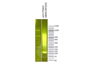
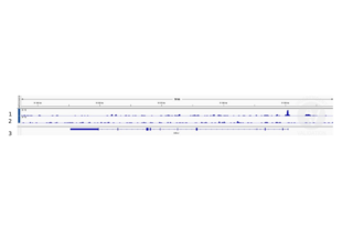
Passed. The anti-FOXO1 antibody ABIN1387620 allows for CUT&RUN targeting profiling of FOXO1 in mouse forelimb cells.
- Anticorps primaire
- ABIN1387620
- Anticorps secondaire
- Full Protocol
- Cell harvest and nuclear extraction
- Dissect 3 Fore limbs (11.5 DAC) from RjOrl:SWISS embryos for each sample.
- Dissociate the tissue into single cells in TrypLE for 15 min at 37 °C.
- Centrifuge cell solution 5 min at 800 x g at RT.
- Remove the liquid carefully.
- Gently resuspend cells in 1 mL of Nuclear Extraction Buffer (20 mM HEPES-KOH pH 8.2, 20% Glycerol, 0,05% IGEPAL, 0.5 mM Spermidine, 10 mM KCl, Roche Complete Protease Inhibitor EDTA-free).
- Move the solution to a 2 mL centrifuge tube.
- Pellet the nuclei 800 x g for 5 min.
- Repeat the NE wash twice for a total of three washes.
- Resuspend the nuclei in 20 µL NE Buffer per sample.
- Concanavalin A beads preparation
- Prepare one 2 mL microcentrifuge tube.
- Gently resuspend the magnetic Concanavalin A Beads (antibodies-online, ABIN6952467).
- Pipette 20 µL Con A Beads slurry for each sample into the 2 mL microcentrifuge tube.
- Place the tube on a magnet stand until the fluid is clear. Remove the liquid carefully.
- Remove the microcentrifuge tube from the magnetic stand.
- Pipette 1 mL Binding Buffer (20 mM HEPES pH 7.5, 10 mM KCl, 1 mM CaCl2, 1 mM MnCl2) into the tube and resuspend ConA beads by gentle pipetting.
- Spin down the liquid from the lid with a quick pulse in a table-top centrifuge.
- Place the tubes on a magnet stand until the fluid is clear. Remove the liquid carefully.
- Remove the microcentrifuge tube from the magnetic stand.
- Repeat the wash twice for a total of three washes.
- Gently resuspend the ConA Beads in a volume of Binding Buffer corresponding to the original volume of bead slurry, i.e. 20 µL per sample.
- Nuclei immobilization – binding to Concanavalin A beads
- Carefully vortex the nuclei suspension and add 20 µL of the Con A beads in Binding Buffer to the cell suspension for each sample.
- Close tube tightly incubates 10 min at 4 °C.
- Put the 1.5 mL tube on the magnet rack and when the liquid is clear remove the supernatant.
- Resuspend the beads in 1 mL of EDTA Wash Buffer (20 mM HEPES pH 7.5, 150 mM NaCl, 0.5 mM Spermidine, Roche Complete Protease Inhibitor EDTA-free, 2 mM EDTA).
- Incubate for 5 min at RT.
- Place the tube on the magnet stand and when the liquid is clear remove the supernatant.
- Resuspend the beads in 200 µL of Wash Buffer (20 mM HEPES pH 7.5, 150 mM NaCl, 0.5 mM Spermidine, Roche Complete Protease Inhibitor EDTA-free) per sample.
- Primary antibody binding
- Divide nuclei suspension into separate 200 µL PCR tubes, one for each antibody (150,000 cells per sample).
- Add 2 µL antibody (anti-FOXO1 antibody ABIN1387620, anti-H3K4me positive control antibody ABIN3023251, guinea pig anti-rabbit IgG negative control antibody ABIN101961) to the respective tube, corresponding to a 1:100 dilution.
- Incubate ON at 4 °C.
- Place the tubes on a magnet stand until the fluid is clear. Remove the liquid carefully.
- Remove the microcentrifuge tubes from the magnetic stand.
- Wash with 200 µL of Wash buffer (to accelerate the process use a multichannel pipette).
- Repeat the wash for a total of five washes.
- pAG-MNase Binding
- Prepare a 1.5 mL microcentrifuge tube containing 200 µL of pAG mix pear sample (200 µL of wash buffer + 120 ng pAG-MNase per sample).
- Place the PCR tubes with the sample on a magnet stand until the fluid is clear. Remove the liquid carefully.
- Remove tubes from the magnetic stand.
- Resuspend the beads in 200 µL of pAG-MNase premix.
- Incubate for 30 min at 4 °C.
- Place the tubes on a magnet stand until the fluid is clear. Remove the liquid carefully.
- Remove the microcentrifuge tubes from the magnetic stand.
- Wash with 200 µL of Wash Buffer using a multichannel pipette to accelerate the process.
- Repeat the wash for a total of five washes.
- Resuspend in 200 µL of Wash Buffer.
- MNase digestion and release of pAG-MNase-antibody-chromatin complexes
- Place PCR tubes on ice and allow to chill.
- Prepare a 1.5 mL microcentrifuge tube with 51 µL of 2 mM CaCl2 mix per sample (50 µL Wash Buffer + 1 µL 100 mM CaCl2) and let it chill on ice.
- Always in ice, place the samples on the magnetic rack and when the liquid is clear remove the supernatant.
- Resuspend the samples in 50 µL of the 2 mM CaCl2 mix and incubate in ice for exactly 30 min.
- Place the sample on the magnet stand and when the liquid is clear move the supernatant in fresh collection tubes with 3 µL of EDTA/EGTA 0.25 M (Digestion buffer).
- Resuspend the sample in 47 µL of 1x Urea STOP Buffer (8.5 M Urea, 100 mM NaCl, 2 mM EGTA, 2 mM EDTA, 0,5% IGEPAL).
- Incubate the samples for 1 h at 4 °C.
- Transfer the supernatant containing the pAG-MNase-bound digested chromatin fragments to the previously collected digestion buffer.
- DNA Clean up
- Take the Mag-Bind® TotalPure NGS beads (Omega Bio-Tek, M1378-01) from the storage and wait until they are RT.
- Add 2x volume of beads to each sample (e.g. 100 µL of beads for 50 µL of sample).
- Incubate the beads and the sample for 15 min at RT.
- During incubation prepare fresh EtOH 80%.
- Place the PCR tubes on a magnet stand and when the liquid is clear remove the supernatant.
- Add 200 µl of fresh 80% EtOH to the sample without disturbing the.
- Incubate 30 sec at RT.
- Remove the EtOH from the sample.
- Repeat the wash with 80% EtOH.
- Resuspend the beads in 25 µL of 10 mM Tris.
- Incubate the sample for 2 min at RT.
- Repeat the 2x beads clean up as described before (this time with 50 µL of beads for each sample).
- Resuspend the beads and DNA in 20 µL of 10 mM Tris.
- Library preparation and sequencing
- Prepare Libraries using KAPA HyperPrep Kit using KAPA Dual-Indexed adapters according to protocol.
- Sequence samples on an Illumina NextSeq 500 sequencer, using a NextSeq 500/550 High Output Kit v2.5 (75 Cycles), 36 bp PE.
- Peak calling
- Trim reads using using bbTools bbduk (BBMap - Bushnell B. - sourceforge.net/projects/bbmap/) to remove adapters, artifacts and repeat sequences.
- Map aligned reads to the mm10 mouse genome using bowtie with options -m 1 -v 0 -I 0 -X 500.
- Use SAMtools to convert SAM files to BAM files and remove duplicates.
- Use BEDtools genomecov to produce Bedgraph files.
- Call peaks using SEACR with a 0.001 threshold and the option norm stringent.
- Notes
The protocol is published in Zambanini, G. et al. A New CUT&RUN Low Volume-Urea (LoV-U) protocol uncovers Wnt/β-catenin tissue-specific genomic targets. Development (2022). PMID 36355069
Validation #104385 (Cleavage Under Targets and Release Using Nuclease)![Testé avec succès 'Independent Validation' signe]()
![Testé avec succès 'Independent Validation' signe]() Validation ImagesProtocole
Validation ImagesProtocole -
- Format
- Liquid
- Concentration
- 1 μg/μL
- Buffer
- 0.01M TBS( pH 7.4) with 1 % BSA, 0.02 % Proclin300 and 50 % Glycerol.
- Agent conservateur
- ProClin
- Précaution d'utilisation
- This product contains ProClin: a POISONOUS AND HAZARDOUS SUBSTANCE, which should be handled by trained staff only.
- Stock
- 4 °C,-20 °C
- Stockage commentaire
- Shipped at 4°C. Store at -20°C for one year. Avoid repeated freeze/thaw cycles.
- Date de péremption
- 12 months
-
- Antigène
- FOXO1 (Forkhead Box O1 (FOXO1))
- Autre désignation
- FOXO1A (FOXO1 Produits)
- Synonymes
- anticorps FKH1, anticorps FKHR, anticorps FOXO1A, anticorps FoxO1a.1, anticorps foxo1, anticorps zgc:153388, anticorps Fkhr, anticorps Foxo1a, anticorps AI876417, anticorps Afxh, anticorps Fkhr1, anticorps fkh1, anticorps fkhr, anticorps foxo1a, anticorps xFoxO1, anticorps FoxO1A, anticorps foxo1a.2, anticorps forkhead box O1, anticorps forkhead box O1 a, anticorps forkhead box O1 L homeolog, anticorps forkhead box O1 b, anticorps FOXO1, anticorps foxo1a, anticorps Foxo1, anticorps foxo1, anticorps foxo1.L, anticorps foxo1b
- Sujet
-
Synonyms: Forkhead box protein O1, Forkhead box protein O1A, Forkhead in rhabdomyosarcoma, FOXO1, FKHR, FOXO1A
Background: Transcription factor that is the main target of insulin signaling and regulates metabolic homeostasis in response to oxidative stress. Binds to the insulin response element (IRE) with consensus sequence 5'-TT[G/A]TTTTG-3' and the related Daf-16 family binding element (DBE) with consensus sequence 5'-TT[G/A]TTTAC-3'. Activity suppressed by insulin. Main regulator of redox balance and osteoblast numbers and controls bone mass. Orchestrates the endocrine function of the skeleton in regulating glucose metabolism. Acts syngernistically with ATF4 to suppress osteocalcin/BGLAP activity, increasing glucose levels and triggering glucose intolerance and insulin insensitivity. Also suppresses the transcriptional activity of RUNX2, an upstream activator of osteocalcin/BGLAP. In hepatocytes, promotes gluconeogenesis by acting together with PPARGC1A to activate the expression of genes such as IGFBP1, G6PC and PPCK1. Important regulator of cell death acting downstream of CDK1, PKB/AKT1 and SKT4/MST1. Promotes neural cell death. Mediates insulin action on adipose. Regulates the expression of adipogenic genes such as PPARG during preadipocyte differentiation and, adipocyte size and adipose tissue-specific gene expression in response to excessive calorie intake. Regulates the transcriptional activity of GADD45A and repair of nitric oxide-damaged DNA in beta-cells.
- Poids moléculaire
- 72kDa
- ID gène
- 2308
- Pathways
- Signalisation PI3K-Akt, Cycle Cellulaire, Fc-epsilon Receptor Signaling Pathway, EGFR Signaling Pathway, Neurotrophin Signaling Pathway, Carbohydrate Homeostasis, Chromatin Binding, Regulation of Carbohydrate Metabolic Process, CXCR4-mediated Signaling Events, BCR Signaling
-


 (1 validation)


