CD200R1L anticorps (AA 151-250)
-
- Antigène Voir toutes CD200R1L Anticorps
- CD200R1L (CD200 Receptor 1-Like (CD200R1L))
-
Épitope
- AA 151-250
-
Reactivité
- Humain
-
Hôte
- Lapin
-
Clonalité
- Polyclonal
-
Conjugué
- Cet anticorp CD200R1L est non-conjugé
- Application
- Western Blotting (WB), ELISA, Flow Cytometry (FACS), Immunofluorescence (Cultured Cells) (IF (cc)), Immunofluorescence (Paraffin-embedded Sections) (IF (p)), Immunohistochemistry (Frozen Sections) (IHC (fro)), Immunohistochemistry (Paraffin-embedded Sections) (IHC (p)), Immunocytochemistry (ICC)
- Réactivité croisée
- Humain
- Purification
- Purified by Protein A.
- Immunogène
- KLH conjugated synthetic peptide derived from human CD200R2
- Isotype
- IgG
-
-
- Indications d'application
-
WB 1:300-5000
ELISA 1:500-1000
FCM 1:20-100
IHC-P 1:200-400
IHC-F 1:100-500
IF(IHC-P) 1:50-200
IF(IHC-F) 1:50-200
IF(ICC) 1:50-200
ICC 1:100-500 - Restrictions
- For Research Use only
-
- by
- Department of Pathology and Molecular Medicine, McMaster University
- No.
- #101354
- Date
- 01.06.2017
- Antigène
- CD200R1L
- Numéro du lot
- 9A13M60
- Application validée
- Immunohistochemistry
- Contrôle positif
- Placental villus from 7w gestation karyotype-normal pregnancy
- Contrôle négative
- KLH antibody ABIN401183
- Conclusion
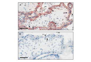
- Passed. ABIN1715098 specifically labels the CD200R1L in human placental villus trophoblast tissue in IHC.
- Anticorps primaire
- ABIN1715098
- Anticorps secondaire
- Dako EnVision-HRP anti-rabbit polymer (Dako, K4003, lot 10095566)
- Full Protocol
- Human placental tissue is fixed in 10% buffered formalin for 24h at RT and embedded in paraffin.
- Cut paraffin blocks with a Leica 12M2255 Microtome into 4µm sections.
- Affix sections to positively charged slides and air dry.
- Deparaffinize and rehydrate sections through graded xylene and graded alcohol series:
- xylene 3x 5 min.
- 100% ethanol 3x 1min.
- 95% ethanol for 1min.
- 70% ethanol for 1min.
- dH2O for 2min.
- Heat the sections in the decloaking Digital Decloaking chamber (Biacore, DC-2002) using factory settings (125°C for 30sec and cool) in 1mM EDTA buffer pH8.0 (0.37g EDTA (disodium salt) in 1l dH2O, adjust to pH8.0 using 1 M NaOH, add 1 small crystal thymol).
- Rinse sections 3 times with 0.05 M Tris-buffered saline pH 7.6 with 0.1% Tween 20 (TBST) 3x for 5min.
- Block sections in 5% normal goat serum (Jackson Immuno Research, 005-000-121, lot 116831) in TBST fo 30min at RT.
- Blot excess serum from sections.
- Incubate sections with
- primary rabbit anti-CD200 Receptor 1-Like (CD200R1L) (AA 150-200) antibody(antibodies-online, ABIN1715098, lot 9A13M60) diluted 1:250 in antibody diluent (Thermo Fisher TA-125-ADQ Lot ADQ 151111) ON at 4°C.
- primary rabbit anti-KLH antibody also antigen-affinity purified (antibodies-online, ABIN401183, lot 30477) diluted 1:250 in Antibody Diluent (as above) ON at 4°C.
- Rinse sections 3x 5min with TBST.
- Stain sections with Dako EnVision-HRP anti-rabbit polymer (Dako, K4003, lot 10095566) for 30min at RT.
- Rinse sections 3x 5min with TBST.
- Stain 30min with AEC (5 ml AEC stock (80mg AEC in 20ml Dimethylformamide), 95 ml 0.05M acetate buffer pH 5.0, filter and add 2 drops of 30% hydrogen peroxide to activate).
- Rinse with pH5.0 acetate buffer.
- Counterstain 1min with 50% Mayer’s hematoxylin.
- Wash in tap water and blue 1min in TBST.
- Mount sections in 15% glycerine/gelatin in TBST pH 8.0 and apply coverslip. Glycerine solution is made with 50 g gelatin (100 bloom) in 150ml TBS pH 8.0, incubated in a 60°C oven until the gelatin is melted, the 175 ml glycerine and a small pellet of thymol is added.
- Allow sections to dry.
- Scan slides using Imagescope and photograph at 400x. Quantification using Image J 1.4.
- Notes
- The result shown is validated by the data of Wang et al. (2014) Acta Obstetrica et Gynecologica Scandinavica where a non-antigen-affinity-purified goat polyclonal antibody to CD200R1 and no negative control antibody was used. Wang et al. confirmed expression of CD200R1 using real time PCR of villus mRNA.
Validation #101354 (Immunohistochemistry)![Testé avec succès 'Independent Validation' signe]()
![Testé avec succès 'Independent Validation' signe]() Validation Images
Validation Images![Representative villus from a 7 week gestation normal pregnancy with “normal karyotype” as determined by PCR testing to exclude trisomy, and digitally photographed at 400x. Staining with CD200R1L antibody ABIN1715098 (A) or KLH antibody ABIN401183 (B). VC: villus core, CT: cytotrophoblast, ST: syncytiotrophoblast, MB: maternal blood space bathing placental villi. Note more intense staining of cytotrophoblasts compared to syncytiotrophoblasts. Bars represent 60µm.]() Representative villus from a 7 week gestation normal pregnancy with “normal karyotype” as determined by PCR testing to exclude trisomy, and digitally photographed at 400x. Staining with CD200R1L antibody ABIN1715098 (A) or KLH antibody ABIN401183 (B). VC: villus core, CT: cytotrophoblast, ST: syncytiotrophoblast, MB: maternal blood space bathing placental villi. Note more intense staining of cytotrophoblasts compared to syncytiotrophoblasts. Bars represent 60µm.
Protocole
Representative villus from a 7 week gestation normal pregnancy with “normal karyotype” as determined by PCR testing to exclude trisomy, and digitally photographed at 400x. Staining with CD200R1L antibody ABIN1715098 (A) or KLH antibody ABIN401183 (B). VC: villus core, CT: cytotrophoblast, ST: syncytiotrophoblast, MB: maternal blood space bathing placental villi. Note more intense staining of cytotrophoblasts compared to syncytiotrophoblasts. Bars represent 60µm.
Protocole - by
- David A. Clark, Department of Medicine and Dept. Pathology and Molecular Medicine, McMaster University
- No.
- #103574
- Date
- 14.12.2018
- Antigène
- CD200R1L
- Numéro du lot
- AD04080113
- Application validée
- Immunohistochemistry
- Contrôle positif
Human term placenta to test anti-CD200R1L
ABIN1715098 lot 9A13M60
- Contrôle négative
Anti-KLH antibody ABIN401183 lot 304770
Diluent
- Conclusion
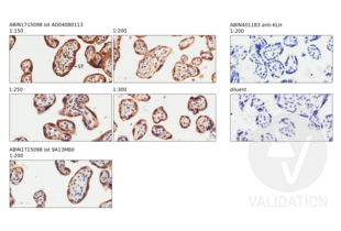
Passed. ABIN1715098 specifically labels CD200R1L in human placental tissue in IHC.
- Anticorps primaire
- ABIN1715098
- Anticorps secondaire
- Bond Polymer Refine Detection kit (Leica, DS9800, lot 62559)
- Full Protocol
- Fix human placental tissue is fixed in 10% buffered formalin for 24h at RT (22-23°C), process and embed in paraffin.
- Cut paraffin blocks with a Leica CM2255 Microtome into 4µm sections.
- Affix sections to positively charged slides and air dry ON at RT.
- Stain slides were stained with a standard IHC-F (with the post primary omitted) to include the following:
- Dewax the slides and hydrate on the automated Leica BOND Rx stainer using Dewax solution (Leica, AR9222).
- Antigen retrieval on a Leica BOND RX automated stainer using epitope retrieval buffer 2 (Leica, AR9640, lot ER20172).
- Stain slides with primary
- rabbit anti-CD200R1L antibody (antibodies-online, ABIN1715098, lot AD04080113) not previously tested,
- rabbit anti- CD200R1L antibody (Antibodies-online, ABIN1715098 lot 9A13M60), or
- rabbit anti-KLH antibody (antibodies-online, ABIN401183, lot 304770)
- diluted 1:200 in Power Vision IHC Super Blocker (Leica, PV6122). The staining protocol incorporates a modified Leica standard protocol IHC-F (with the post-primary step omitted) and uses the standard times outlined in the machine protocol: primary antibody 15min, Polymer 8min, DAB 10min and Hematoxylin 5min.
- The Bond Polymer Refine Detection kit (Leica, DS9800, lot 62559) containing peroxidase block, post primary antibody, Polymer as well as DAB chromogen and hematoxylin counterstain was used as outlined in the standard protocol IHC-F.
- Remove slides from the Leica Bond Rx and then dehydrate in ethanol and clear in xylene.
- Mount slides in Fisher Chemical Permount Mounting Medium (Fisher Sci., SP15-500, lot 162767) and apply coverslip
- Once the slide-coverslip edges are dry, scan the slides using Leica Imagescope (40x) and photograph into jpg files at 400x.
- Notes
Several dilutions of ABIN1715098 lot AD04080113 were tested and optimal staining of villus trophoblasts was obtained at a dilution of 1:200. Staining was obtained at a dilution as low as 1:300, but 1:200 was judged optimal for best detail. Results were comparable to those obtained with the previous lot 9A13M60. ABIN1715098 stained villus trophoblasts in term placenta as described in Clark et al. (2017). ABIN1715098 stained syncytiotrophoblasts, and cytotorophoblasts in placental villi as well as villus stromal macrophage-like Hoffbauer cells. No staining was observed with the negative control
-
- Format
- Liquid
- Concentration
- 1 μg/μL
- Buffer
- 0.01M TBS( pH 7.4) with 1 % BSA, 0.02 % Proclin300 and 50 % Glycerol.
- Agent conservateur
- ProClin
- Précaution d'utilisation
- This product contains ProClin: a POISONOUS AND HAZARDOUS SUBSTANCE, which should be handled by trained staff only.
- Stock
- 4 °C,-20 °C
- Stockage commentaire
- Shipped at 4°C. Store at -20°C for one year. Avoid repeated freeze/thaw cycles.
- Date de péremption
- 12 months
-
-
: "Circulating CD200 is increased in the secretory phase of women with endometriosis as is endometrial mRNA, and endometrial stromal cell CD200R1 is increased in spite of reduced mRNA." dans: American journal of reproductive immunology (New York, N.Y. : 1989), Vol. 89, Issue 1, pp. e13655, (2022) (PubMed).
: "Changes in expression of the CD200 tolerance-signaling molecule and its receptor (CD200R) by villus trophoblasts during first trimester missed abortion and in chronic histiocytic intervillositis." dans: American journal of reproductive immunology (New York, N.Y. : 1989), Vol. 78, Issue 1, (2017) (PubMed).
: "The Receptor for the CD200 Tolerance-Signaling Molecule Associated with Successful Pregnancy is Expressed by Early-Stage Breast Cancer Cells in 80% of Patients and by Term Placental Trophoblasts." dans: American journal of reproductive immunology (New York, N.Y. : 1989), Vol. 74, Issue 5, pp. 387-91, (2015) (PubMed).
: "The CD200 tolerance-signaling molecule and its receptor, CD200R1, are expressed in human placental villus trophoblast and in peri-implant decidua by 5 weeks' gestation." dans: Journal of reproductive immunology, Vol. 112, pp. 20-3, (2015) (PubMed).
-
: "Circulating CD200 is increased in the secretory phase of women with endometriosis as is endometrial mRNA, and endometrial stromal cell CD200R1 is increased in spite of reduced mRNA." dans: American journal of reproductive immunology (New York, N.Y. : 1989), Vol. 89, Issue 1, pp. e13655, (2022) (PubMed).
-
- Antigène
- CD200R1L (CD200 Receptor 1-Like (CD200R1L))
- Autre désignation
- CD200R2 (CD200R1L Produits)
- Synonymes
- anticorps CD200R2, anticorps CD200RLa, anticorps AY230198, anticorps Cd200r1l, anticorps CD200 receptor 1 like, anticorps Cd200 receptor 2, anticorps CD200R1L, anticorps Cd200r2
- Sujet
-
Synonyms: CD200R2, CD200RLa, Cell surface glycoprotein CD200 receptor 2, CD200 cell surface glycoprotein receptor-like 2, CD200 receptor-like 2, HuCD200R2, CD200 cell surface glycoprotein receptor-like a, Cell surface glycoprotein CD200 receptor 1-like, Cell surface glycoprotein OX2 receptor 2, CD200R1L
Background: May be a receptor for the CD200/OX2 cell surface glycoprotein.
- ID gène
- 344807
- UniProt
- Q6Q8B3
-


 Digital jpg photomicrographs at 400x from Imagescope using a human term placenta stained with the ABIN1715098 lot AD04080113 (diluted 1:150, 1:200, 1:250, 1:300) and 9A13M60 (diluted 1:200) and ABIN401183 and the diluent only as negative controls.
VC=placental villus core; CT=cytotrophoblast inner layer; ST=syncytiotrophoblast outer layer.


 (4 références)
 (2 validations)


