beta Actin anticorps (AA 2-16)
-
- Antigène Voir toutes beta Actin (ACTB) Anticorps
- beta Actin (ACTB) (Actin, beta (ACTB))
-
Épitope
- AA 2-16
-
Reactivité
- Souris, Rat, Poulet, Poisson zèbre (Danio rerio)
-
Hôte
- Lapin
-
Clonalité
- Polyclonal
-
Conjugué
- Cet anticorp beta Actin est non-conjugé
-
Application
- Western Blotting (WB), Immunohistochemistry (IHC), Immunocytochemistry (ICC)
- Réactivité croisée (Details)
- May cross-react to alpha- and gamma-actin due to sequence homology.
- Purification
- Affinity purified with the immunogen. Rabbit serum albumin was added for stabilization.
- Immunogène
- Synthetic peptide (aa 2-16 of mouse beta-actin) coupled to KLH via a C-terminally added cysteine.
- Top Product
- Discover our top product ACTB Anticorps primaire
-
-
- Indications d'application
-
WB: 1 : 1000 up to 1 : 5000
IP: not tested yet
ICC: 1 : 200 up to 1 : 500
IHC: 1 : 200 - Commentaires
-
ICC: Methanol fixation is recommended.
- Restrictions
- For Research Use only
-
- by
- Kinexus Bioinformatics Corporation
- No.
- #029836
- Date
- 22.06.2015
- Antigène
- Numéro du lot
- 251003/3
- Application validée
- Western Blotting
- Contrôle positif
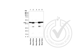
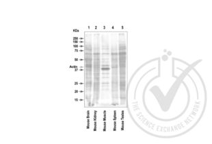
- mouse brain, mouse kidney, mouse muscle, mouse spleen, mouse testes
- Contrôle négative
- none [ubiquitously expressed]
- Conclusion
- A band was observed in the positive controls at the expected size (~42 kDa)
- Anticorps primaire
- Antigen: Beta-Actin, (ACTB) (AA 2-16)
- Catalog number: ABIN1742553
- Lot number: 251003/3
- Dilution: 1:1000
- Anticorps secondaire
- Antibody: Donkey anti-Rabbit IgG Antibody (HRP)
- Lot number: F0613
- Dilution: 1:10,000
- Full Protocol
- 1. Cell/tissue total protein lysates were boiled in 1X SDS Sample Buffer containing 1% SDS and 1.25% β-mercaptoethanol at 95°C for 5 minutes prior to loading.
- 2. 15 μg of boiled lysate were loaded and resolved on a 12% SDS-polyacrylamide gel.
- 3. The Precision Plus Protein™ All Blue Prestained Standards from BioRad (161-0373) were used as molecular mass markers.
- 4. Proteins were transferred onto nitrocellulose membrane by tank transfer and protein transfer was confirmed with Ponceau S staining.
- 5. The immunoblot membrane was blocked in 2.5% skim milk and 1.5% BSA solution in TTBS at room temperature for 60 minutes.
- 6. The membrane was washed in TTBS twice for 5 minutes each.
- 7. The membrane was immersed with the protein side up in the antibody solution in TBS and incubated overnight at 4°C with gentle agitation.
- 8. The membrane was rinsed twice with TTBS.
- 9. The membrane was washed in TTBS twice for 5 minutes each.
- 10. The membrane was washed in TTBS once for 15 minutes.
- 11. The membrane was incubated in the HRP-conjugated secondary antibody solution in TBS for 60 minutes at room temperature with gentle agitation.
- 12. The membrane was rinsed twice with TTBS.
- 13. The membrane was washed in TTBS twice for 5 minutes each.
- 14. The membrane was washed in TTBS once for 15 minutes.
- 15. Signals were detected by chemiluminescence (ECL). The blot was scanned for 160 seconds.
- 16. The membrane was rinsed three times with TTBS.
- 17. Incubated in Acidic Glycine Stripping Buffer at room temperature with gentle agitation for 3 times, 10 minutes each.
- 18. The membrane was washed in TTBS 3 times for 5 minutes each.
- 19. Repeated Steps 4-14 with the loading control antibody (for β3−Tubulin).
- 20. Repeated Steps 4-14 with the loading control antibody and its matching secondary antibody. The blot was scanned for 160 seconds.
- Notes
- A protein band was observed at the expected target size of β−Actin of 42 kDa in all mouse extracts tested. Lower molecular weight bands of approximately 28-30 kDa were also observed in the mouse kidney and spleen tissue samples. Three main groups of actin have been identified. The β-actins and γ-actins co-exist in most cell types as components of the cytoskeleton, whereas the α-actin are found in muscle tissues. In our experience, we find that the level of actin does vary widely between different cell and tissue types.
Validation #029836 (Western Blotting)![Testé avec succès 'Independent Validation' signe]()
![Testé avec succès 'Independent Validation' signe]() Validation Images
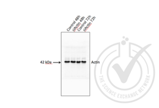
Validation Images![Figure 3. Western blot for beta actin (ACTB) using attempted siRNA silencing. Arrowhead indicates the expected molecular weight of ~42 kDa. HeLa cells were transfected with beta actin siRNA (Catalog number 12935141, Life Technologies) using Lipofectamine 2000, according to manufacturer's instructions. Total cell lysates were made from both floating and adherent cells harvested 48 hours and 72 hours after transfection. siRNA silencing of beta actin was not detected.]() Figure 3. Western blot for beta actin (ACTB) using attempted siRNA silencing. Arrowhead indicates the expected molecular weight of ~42 kDa. HeLa cells were transfected with beta actin siRNA (Catalog number 12935141, Life Technologies) using Lipofectamine 2000, according to manufacturer's instructions. Total cell lysates were made from both floating and adherent cells harvested 48 hours and 72 hours after transfection. siRNA silencing of beta actin was not detected.
Protocole
Figure 3. Western blot for beta actin (ACTB) using attempted siRNA silencing. Arrowhead indicates the expected molecular weight of ~42 kDa. HeLa cells were transfected with beta actin siRNA (Catalog number 12935141, Life Technologies) using Lipofectamine 2000, according to manufacturer's instructions. Total cell lysates were made from both floating and adherent cells harvested 48 hours and 72 hours after transfection. siRNA silencing of beta actin was not detected.
Protocole -
- Format
- Lyophilized
- Reconstitution
- For reconstitution add 100 µL H2O to get a 1mg/ml solution of antibody in PBS. Then aliquot and store at -20 °C until use.
- Buffer
- PBS
- Conseil sur la manipulation
- Affinity purified antibodies are less robust than antisera, since protease inhibitors are also removed during purification. Hence, storage at 4 °C for prolonged periods (i.e. several weeks), is not recommended.
- Stock
- -20 °C
- Stockage commentaire
- Unlabeled lyophilized antibodies are stable in this form without loss of quality at ambient temperatures for several weeks or even months. They can be stored at 4°C for several years. Lyophilized antibodies must not be stored in the freezer, they may be destroyed!
-
-
: "Retinoic acid‑incorporated glycol chitosan nanoparticles inhibit the expression of Ezh2 in U118 and U138 human glioma cells." dans: Molecular medicine reports, Vol. 12, Issue 5, pp. 6642-8, (2016) (PubMed).
: "The α-subunit of the trimeric GTPase Go2 regulates axonal growth." dans: Journal of neurochemistry, Vol. 124, Issue 6, pp. 782-94, (2013) (PubMed).
: "Novel application of human neurons derived from induced pluripotent stem cells for highly sensitive botulinum neurotoxin detection." dans: Toxicological sciences : an official journal of the Society of Toxicology, Vol. 126, Issue 2, pp. 426-35, (2012) (PubMed).
-
: "Retinoic acid‑incorporated glycol chitosan nanoparticles inhibit the expression of Ezh2 in U118 and U138 human glioma cells." dans: Molecular medicine reports, Vol. 12, Issue 5, pp. 6642-8, (2016) (PubMed).
-
- Antigène
- beta Actin (ACTB) (Actin, beta (ACTB))
- Autre désignation
- beta-Actin (ACTB Produits)
- Synonymes
- anticorps BRWS1, anticorps PS1TP5BP1, anticorps ACTA1, anticorps Actx, anticorps E430023M04Rik, anticorps beta-actin, anticorps ps1tp5bp1, anticorps act-2, anticorps actb, anticorps B-actin, anticorps LOC100174883, anticorps LOC100304816, anticorps A, anticorps ACT5C, anticorps Ac5C, anticorps Act, anticorps Act-5C, anticorps Act5, anticorps Act5c, anticorps Actin, anticorps Actin/BAP47, anticorps Actin5C, anticorps BAP47, anticorps Bap47, anticorps CG4027, anticorps Dmel\\CG4027, anticorps M32055, anticorps T11, anticorps act, anticorps act 5C, anticorps act5C, anticorps actin, anticorps actin5C, anticorps anon-EST:fe2D2, anticorps beta-actin/Bap47, anticorps cyt5C, anticorps l(1)G0009, anticorps l(1)G0010, anticorps l(1)G0025, anticorps l(1)G0079, anticorps l(1)G0117, anticorps l(1)G0177, anticorps l(1)G0245, anticorps l(1)G0330, anticorps l(1)G0420, anticorps l(1)G0486, anticorps Bact, anticorps ACT-5, anticorps ACTB, anticorps B-ACTZF, anticorps actba, anticorps bact, anticorps bactin1, anticorps bactzf, anticorps wu:fd18f05, anticorps A26C1B, anticorps POTE2alpha, anticorps POTEACTIN, anticorps Beta-actin, anticorps actin beta, anticorps actin, beta, anticorps cytoplasmic actin OlCA1, anticorps beta-actin, anticorps Actin 5C, anticorps actin gamma 1, anticorps actin, beta 1, anticorps actin, beta L homeolog, anticorps beta actin, anticorps POTE ankyrin domain family member F, anticorps actin, beta like 2, anticorps actin, cytoplasmic 1, anticorps ACTB, anticorps Actb, anticorps actb, anticorps LOC100174883, anticorps LOC100304816, anticorps Act5C, anticorps ACTG1, anticorps actb1, anticorps actb.L, anticorps LOC100136352, anticorps POTEF, anticorps ACTBL2, anticorps LOC109073280
- Pathways
- Myometrial Relaxation and Contraction, Cell-Cell Junction Organization, Maintenance of Protein Location, Phototransduction
-


 (3 références)
 (1 validation)


