C9 anticorps (Center)
-
- Antigène Voir toutes C9 Anticorps
- C9 (Complement Component C9 (C9))
-
Épitope
- AA 191-220, Center
-
Reactivité
- Humain
-
Hôte
- Lapin
-
Clonalité
- Polyclonal
-
Conjugué
- Cet anticorp C9 est non-conjugé
-
Application
- Western Blotting (WB), Immunohistochemistry (Paraffin-embedded Sections) (IHC (p))
- Specificité
- This C9 antibody is generated from rabbits immunized with a KLH conjugated synthetic peptide between 190-220 amino acids from the Central region of human C9.
- Purification
- This antibody is purified through a protein A column, followed by peptide affinity purification.
- Immunogène
- This C9 antibody is generated from rabbits immunized with a KLH conjugated synthetic peptide between 191-220 AA from the Central region of human C9.
- Clone
- RB33918
- Isotype
- Ig Fraction
-
-
- Indications d'application
- WB = 1:1000, IHC (p) = 1:10-50
- Restrictions
- For Research Use only
-
- by
- Protein research group, Department of Biochemistry and Molecular Biology, University of Southern Denmark, Odense Denmark
- No.
- #102069
- Date
- 17.01.2018
- Antigène
- C9
- Numéro du lot
- Application validée
- Western Blotting
- Contrôle positif
- Human endothelial cells with nanoparticle treatment
- Contrôle négative
- Human endothelial cells and astrocytes without nanoparticle treatment
- Conclusion
Passed. ABIN657704 specifically recognizes C9 in human endothelial cells.
- Anticorps primaire
- ABIN657704
- Anticorps secondaire
- anti-rabbit IgG HRP-linked antibody (Cell Signaling, 7074S)
- Full Protocol
- Grow human endothelial cells and astrocytes in EndoGRO-MV Complete Media kit (Millipore, SCME004, lot 2885245) supplemented with serum (Millipore, lot 1000958) at 37°C and 5% CO2 in 8ml on a petri dish to 80-90 % confluency.
- Add PVP coated silver nanoparticles (50nm Silver Nanospheres, Nanocomposix USA lot no JRC0234) to the endothelial cells for 24h and 48h.
- Lyse 106 cells in 100µl per well cold lysis buffer (6M Urea, 2M Thiourea, 10mM DTT).
- Determine total protein content of the lysates using Qubit Protein Assay Kit (ThermoFisher Scientific, Q33211).
- Denature 20µg of total protein for 5min at 95°C in 5µl Pierce LDS Sample Buffer, Non-Reducing (Thermo Scientific, b31010, lot 151109002) and separate proteins in an XCell SureLock Mini-Cell (Life Technologies, EI0001, lot 007469934) in a Bolt 4-12% Bis-Tris Plus gel (Thermo scientific, NW04125BOX, lot 17120471) for 60min at 100V.
- Transfer proteins onto PVDF membrane (Sigma-Aldrich, IPFL00005) with a Western blotting system for 20min at 18V.
- Block the membrane with TBST and 5% non-fat milk powder (Sigma-Aldrich, 70166-500G, lot SZBF3420V) for 1h at RT.
- Incubation with primary rabbit anti-C9 antibody (antibodies-online, ABIN657704, lot SA110711AL) diluted 1:1000 TBST and 5% non-fat milk powder for ON at 4°C.
- Wash membrane 3x for 15min with TBST.
- Incubation with secondary anti-rabbit IgG HRP-linked antibody (Cell Signaling, 7074S) diluted 1:10000 in TBST and 5% non-fat milk powder for 2h at RT.
- Wash membrane 3x for 15min with TBS.
- Reveal protein bands using Luminata Forte Western HRP Substrate (Millipore, WBLUF0100) on a UVP multispectral imaging system (AH diagnostics, Denmark).
- Incubate the membrane in Restore Plus Western blot Stripping Buffer (Thermo Scientific, 46430, lot RL241559) for 20min at RT.
- Wash membrane 3x for 15min with TBST.
- Incubation with rabbit anti-GAPDH antibody (Cell Signaling Technology, 2118) diluted 1:5000 in TBST and 5% non-fat milk powder for 2h at RT.
- Wash membrane 3x for 15min with TBS.
- Reveal protein bands as described above.
- Notes
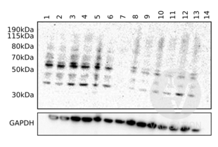
Upon addition of the nanoparticles, the endothelial cells’ proteome revealed high expression of C9 at the 24h time point and reduced expression at the 48h time point. We validated the proteomics results by western blotting and we confirm that the C9 antibody ABIN657704 reveals a protein of the expected molecular weight (60kDa) in lysates of endothelial cells. The protein band is only visible in the nanoparticle treated cells but not the negative controls.
Validation #102069 (Western Blotting)![Testé avec succès 'Independent Validation' signe]()
![Testé avec succès 'Independent Validation' signe]() Validation Images
Validation Images![Immunoblot using ABIN657704 on human endothelial cell and human astrocytes lysates. C9 protein band intensity is increased in the human endothelial cell after 24h (lane 1-6) compared to 48h (lane 8-13) when treated with nanoparticles. Negative controls are endothelial cells (lane 7) and astrocytes (lane 14) without nanoparticle treatment.]() Immunoblot using ABIN657704 on human endothelial cell and human astrocytes lysates. C9 protein band intensity is increased in the human endothelial cell after 24h (lane 1-6) compared to 48h (lane 8-13) when treated with nanoparticles. Negative controls are endothelial cells (lane 7) and astrocytes (lane 14) without nanoparticle treatment.
Protocole
Immunoblot using ABIN657704 on human endothelial cell and human astrocytes lysates. C9 protein band intensity is increased in the human endothelial cell after 24h (lane 1-6) compared to 48h (lane 8-13) when treated with nanoparticles. Negative controls are endothelial cells (lane 7) and astrocytes (lane 14) without nanoparticle treatment.
Protocole -
- Format
- Liquid
- Concentration
- 0.36 mg/mL
- Buffer
- PBS with 0.09 % (W/V) sodium azide
- Agent conservateur
- Sodium azide
- Précaution d'utilisation
- This product contains sodium azide: a POISONOUS AND HAZARDOUS SUBSTANCE which should be handled by trained staff only.
- Stock
- 4 °C/-20 °C
- Stockage commentaire
- C9 Antibody (Center) can be refrigerated at 2-8 °C for up to 6 months. For long term storage, place the at -20 °C.
- Date de péremption
- 6 months
-
- Antigène
- C9 (Complement Component C9 (C9))
- Autre désignation
- C9 (C9 Produits)
- Synonymes
- anticorps LOC100037951, anticorps LOC100136130, anticorps c9, anticorps wu:fb60b05, anticorps wu:fd50e04, anticorps zgc:112272, anticorps complement C9, anticorps complement component 9, anticorps complement component 9 L homeolog, anticorps C9, anticorps c9, anticorps c9.L
- Sujet
-
This gene encodes the final component of the complement system. It participates in the formation of the Membrane Attack Complex (MAC). The MAC assembles on bacterial membranes to form a pore, permitting disruption of bacterial membrane organization. Mutations in this gene cause component C9 deficiency. [provided by RefSeq].
Synonyms: Complement component C9,C9, - Poids moléculaire
- 63173 DA
- ID gène
- 735
- NCBI Accession
- NP_001728
- UniProt
- P02748
- Pathways
- Système du Complément
-


 (1 validation)


