TJP1 anticorps (AA 1551-1702)
-
- Antigène Voir toutes TJP1 Anticorps
- TJP1 (Tight Junction Protein 1 (TJP1))
-
Épitope
- AA 1551-1702
-
Reactivité
- Humain, Souris, Rat, Porc, Boeuf (Vache)
-
Hôte
- Lapin
-
Clonalité
- Polyclonal
-
Conjugué
- Cet anticorp TJP1 est non-conjugé
-
Application
- Western Blotting (WB), ELISA, Flow Cytometry (FACS), Immunohistochemistry (Paraffin-embedded Sections) (IHC (p)), Immunofluorescence (Paraffin-embedded Sections) (IF (p)), Immunofluorescence (Cultured Cells) (IF (cc)), Immunohistochemistry (Frozen Sections) (IHC (fro))
- Réactivité croisée
- Boeuf (Vache), Humain, Souris, Porc, Rat
- Homologie
- Dog,Cow,Chicken,Rabbit,Guinea Pig
- Purification
- Purified by Protein A.
- Immunogène
- KLH conjugated synthetic peptide derived from human ZO-1
- Isotype
- IgG
-
-
- Indications d'application
-
WB 1:300-5000
ELISA 1:500-1000
FCM 1:20-100
IHC-P 1:200-400
IHC-F 1:100-500
IF(IHC-P) 1:50-200
IF(IHC-F) 1:50-200
IF(ICC) 1:50-200 - Restrictions
- For Research Use only
-
- by
- Immunohistochemistry Core, NYU Langone
- No.
- #029577
- Date
- 18.01.2014
- Antigène
- Numéro du lot
- 130913
- Application validée
- Immunohistochemistry
- Contrôle positif
- Testis
- Contrôle négative
- See Controls section for negative controls
- Conclusion
- Signal was detected in positive control tissue, and not detected in a tissue expected to express very low levels of the target antigen.
- Anticorps primaire
- Antibody: human Tight Junction Protein 1 (Zona Occludens 1) (TJP1)
- Catalog number: ABIN675024
- Lot number: 130913
- Anticorps secondaire
- Antibody: Biotinylated goat anti-rabbit/anti-mouse (Kit)
- Lot number: D05923BA
- Full Protocol
- Immunohistochemistry was performed on a Ventana NexES automated platform, instrument manufacturer specific reagents are italicized.
- 1. Slides were preheated in convection oven at 60°C for 30 minutes
- 2. Deparaffinization procedure: - 3 changes of Xylene, 5 minutes each - 3 changes of 100% Ethanol, 3 minutes each - 3 changes of 95% Ethanol, 3 minutes each - Rinsed in distilled water, 3 changes
- 1. Heat retrieval procedure - Slides retrieved in 10.0 mM Citrate, pH6.0 in a 1000W microwave oven (~100°C) for 15 minutes. - Slides were allowed to cool (in citrate) for 30 minutes. - Slides were washed x 3 in Distilled water
- 1. NexES instrument procedure, iVIEW DAB paraffin protocol (*abridged*): - Slide chamber warmed to 37°C
- 1. Slides rinsed with *reaction buffer* x 3
- 1. *iVIEW Inhibitor (H2O2)* applied and incubated for 4 minutes
- 1. Slides rinsed with *reaction buffer*
- 1. Antibody Application - Primary antibody diluted 1:250 in PBS (100 microliters applied/slide) - Ventana Isotype control applied neat - Slides incubated overnight at room temperature (~12 hours ~25°C)
- 1. Slides rinsed with *reaction buffer* x3
- 1. *iVIEW Biotinylated IgG* applied and incubated for 8 minutes
- 1. Slides rinsed with *reaction buffer*
- 1. *iVIEW Streptavidin-Horseradish Peroxidase* applied and incubated for 8 minutes
- 1. Slides rinsed with *reaction buffer*
- 1. *iVIEW DAB/H2O2* applied and incubated for 8 minutes
- 1. Slides rinsed with *reaction buffer*
- 1. *iVIEW Copper* applied and incubated for 4 minutes
- 1. Slides rinsed with *reaction buffer*
- 1. Slides washed in Dawn Detergent/tap water
- 1. Counterstain Procedure - Hematoxylin (Leica 560 MX) 30 seconds - Slides washed in tap water, 1 minute - Decolorized (10% Acetic Acid in 70% ethanol), 1 minute - Slides washed in tap water, 1 minute - Bluing (Austin Clear Ammonia), 1 minute - Slides washed in tap water, 1 minute
- 1. Dehydration/coverslipping procedure: - 3 changes of 95% Ethanol, 3 minutes each - 3 changes of 100% Ethanol, 3 minutes each - 3 changes of Xylene, 5 minutes each - Mounted with Permount
- 1. Imaging: Leica SCN 400F Whole Slide Scanner with Digital Image Hub and Leica Slidepath software
- Notes
- Deviations from protocol/procedure supplied by manufacturer (attached).
- Step 1: Heated tissue 60°C for 30 minutes; manufacturer heats for 45 minutes.
- Step 2: No ethanol wash was performed during deparaffinization; manufacturer includes 1 wash of 80% ethanol for 3 minutes.
- Step 3.1: Slides were heated for 15 minutes; manufacturer provides a range of 15-20 minutes.
- Step 3.2: Slides were cooled for 30 minutes; manufacturer cools for 20 minutes.
- Step 4: Italicized reagents and incubation time are fixed instrument parameters.
- Step 5: Secondary species-specific serum block not used; manufacturer blocks with 5% normal goat serum for 2 hours.
- Step 8.1: Antibody diluted in PBS at 1:250; manufacture did not recommend diluent or dilution.
- Step 8.2.1: Primary antibody incubated at room temperature overnight; manufacturer incubates overnight 4°C with agitation.
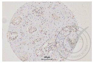
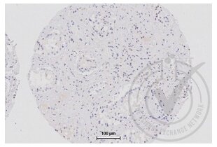
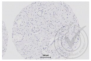
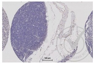
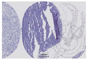
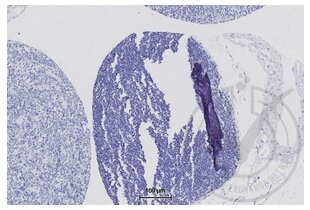
- Tissue Interpretation (limited): - TJP1: Under the staining parameters described above, testis stained weakly (ducts) positive (Figure 1). Substantial signal detected in limited number of other tissues, including: breast, normal (NOS); pancreatic cancer (NOS), and stomach, normal (NOS). Most tissues showed low level of specific signal. Thymus did not have any detectable signal (Figure 4).
- I-NC (Isotype negative control): No signal detected
- B-NC (Blank negative control): No signal detected
- Signal Localization: - Signal to noise was adequate with cytoplasmic, nuclear subcellular localization observed. Rare inner-membrane and no distinct membrane signal observed.
Validation #029577 (Immunohistochemistry)![Testé avec succès 'Independent Validation' signe]()
![Testé avec succès 'Independent Validation' signe]() Validation ImagesProtocole
Validation ImagesProtocole -
- Format
- Liquid
- Concentration
- 1 μg/μL
- Buffer
- 0.01M TBS( pH 7.4) with 1 % BSA, 0.02 % Proclin300 and 50 % Glycerol.
- Agent conservateur
- ProClin
- Précaution d'utilisation
- This product contains ProClin: a POISONOUS AND HAZARDOUS SUBSTANCE, which should be handled by trained staff only.
- Stock
- 4 °C,-20 °C
- Stockage commentaire
- Shipped at 4°C. Store at -20°C for one year. Avoid repeated freeze/thaw cycles.
- Date de péremption
- 12 months
-
-
: "Rhein ameliorates lipopolysaccharide-induced intestinal barrier injury via modulation of Nrf2 and MAPKs." dans: Life sciences, Vol. 216, pp. 168-175, (2019) (PubMed).
: "The Superantigen Toxic Shock Syndrome Toxin-1 Alters Human Aortic Endothelial Cell Function." dans: Infection and immunity, (2018) (PubMed).
: "Huangqin-tang ameliorates dextran sodium sulphate-induced colitis by regulating intestinal epithelial cell homeostasis, inflammation and immune response." dans: Scientific reports, Vol. 6, pp. 39299, (2018) (PubMed).
: "?-Conglycinin reduces the tight junction occludin and ZO-1 expression in IPEC-J2." dans: International journal of molecular sciences, Vol. 15, Issue 2, pp. 1915-26, (2014) (PubMed).
: "Chlorogenic acid decreases intestinal permeability and increases expression of intestinal tight junction proteins in weaned rats challenged with LPS." dans: PLoS ONE, Vol. 9, Issue 6, pp. e97815, (2014) (PubMed).
: "Effects of soybean agglutinin on intestinal barrier permeability and tight junction protein expression in weaned piglets." dans: International journal of molecular sciences, Vol. 12, Issue 12, pp. 8502-12, (2012) (PubMed).
: "Calcium-activated potassium channel activator down-regulated the expression of tight junction protein in brain tumor model in rats." dans: Neuroscience letters, Vol. 493, Issue 3, pp. 140-4, (2011) (PubMed).
-
: "Rhein ameliorates lipopolysaccharide-induced intestinal barrier injury via modulation of Nrf2 and MAPKs." dans: Life sciences, Vol. 216, pp. 168-175, (2019) (PubMed).
-
- Antigène
- TJP1 (Tight Junction Protein 1 (TJP1))
- Autre désignation
- ZO-1 (TJP1 Produits)
- Synonymes
- anticorps ZO-1, anticorps ZO1, anticorps zo1, anticorps tight junction protein 1, anticorps tight junction protein ZO-1, anticorps TJP1, anticorps Tjp1, anticorps LOC396567
- Sujet
-
Synonyms: ZO-1, Tight junction protein ZO-1, Tight junction protein 1, Zona occludens protein 1, Zonula occludens protein 1, TJP1, ZO1
Background: The N-terminal may be involved in transducing a signal required for tight junction assembly, while the C-terminal may have specific properties of tight junctions. The alpha domain might be involved in stabilizing junctions. Plays a role in the regulation of cell migration by targeting CDC42BPB to the leading edge of migrating cells.
- ID gène
- 7082
- UniProt
- Q07157
- Pathways
- Carbohydrate Homeostasis, Cell-Cell Junction Organization
-

 (7 références)
 (1 validation)